A strep throat in childhood could scar a heart valve, and twenty years later the extra blood volume of pregnancy was usually the first thing to find the damage it left.
Rheumatic heart disease is an illness of children that presents its bill to adults. The fever comes at eight or ten; the valve it scars works quietly for twenty years.
From Throat to Valve
The World Health Organization describes the mechanism plainly: “The disease results from damage to heart valves caused by one or several episodes of rheumatic fever, an autoimmune inflammatory reaction to throat infection caused by group A streptococci.” The infection is an ordinary sore throat. The reaction that follows is the body’s own, and it can leave the mitral valve — the door between the left atrium and the left ventricle — thickened, fused and narrowed. Rheumatic fever, the organization notes, “mostly affects children and adolescents.” The narrowing is called mitral stenosis, and for years it produces nothing a patient would report. A doctor listening carefully can hear it. Nobody listens carefully to a well twenty-year-old.
Why Pregnancy Found It
Pregnancy is the test. The European Society of Cardiology is direct: “During pregnancy, there is a progressive increase in blood volume, heart rate and cardiac output which reach their peak levels at 28-32 weeks of gestation.” The Methodist DeBakey Cardiovascular Journal puts the rise in cardiac output at thirty to fifty per cent above pre-pregnancy levels. A normal valve passes it; a narrowed one cannot. Because mitral stenosis is, in the society’s words, “a fixed obstruction to left ventricular inflow,” the changes of pregnancy “usually cause decompensation of a previously asymptomatic MS.” Heart rate compounds it: a rapid rate “shortens diastolic filling time, increases the left atrial pressure and the pulmonary venous pressure.” The pressure backs up into the lungs, and the woman can no longer lie flat.
Before the Cardiac Clinics
What that cost is recorded in a 2021 history in Global Heart, the journal of the World Heart Federation. The nineteenth-century physician Angus Macdonald reported a series in which nine of fourteen pregnancies in women with rheumatic heart disease were fatal. James Mackenzie recorded finding “a presystolic murmur” in a young woman only after she miscarried. What changed was not a drug. It was a room. The Boston Lying-In Hospital opened a cardiac clinic in 1921, and maternal mortality among its cardiac patients fell from twenty per cent — ninety-three of every hundred of those deaths from rheumatic heart disease — to under five per cent in three years. A Scottish joint cardiac and obstetric clinic founded in 1928 saw cardiac deaths fall from 6.3 per cent to 0.9 per cent by 1947. A cardiologist and an obstetrician looking at the same woman on the same afternoon was the whole intervention.
What 1965 Could Do
By the mid-1960s the disease was retreating; the same history notes that by that decade “the overall prevalence of RHD was waning in high-income countries.” What a physician had to work with was modest: rest, oxygen, salt restriction, cautious diuretics, digitalis if the atrium went into fibrillation, and penicillin to prevent a second attack. Surgery existed. R. C. Brock read a paper titled “Valvotomy in Pregnancy” before the Royal Society of Medicine, published in its Proceedings in August 1952, so operating on a narrowed valve in pregnancy was already under discussion more than a decade earlier. Nothing read for this post sets out what any American hospital’s protocol was in 1965, and this one does not invent one.
Counting the Fetal Heart
The other half of that bedside was the baby, and there the instrument was an ear. A 2021 review in Perinatology dates the first listening to 1818, when Mayor heard fetal heart sounds “through the abdominal wall of a pregnant woman,” and notes that from the 1800s onward rates below 100 or above 160 beats a minute “were suspected indicators of fetal asphyxia” — thresholds still in service a century and a half later. “In 1917,” the review records, “the Delee-Hillis fetal heart rate stethoscope was introduced to the world.” Edward Hon reached the fetus electrically in 1958 with “an electrode that could be directly attached to the fetal scalp.” But continuous electronic monitoring did not become “the primary method for assessing fetal well-being” until the 1970s. In 1965 the record was a number, written once, by hand.
What the Novel Takes
A Stone’s Throw puts a reader at that bedside. Helen Calloway is an intern at Los Angeles County General in 1965 when a consult comes down: a twenty-eight-year-old in a first pregnancy, rheumatic fever in childhood, breathless at rest and unable to lie flat. Helen hears what the history predicts: a low murmur at the apex with a snap before it, fine crackles at both lung bases, a pulse of one hundred and twelve. The cardiologist’s orders are the period’s entire armoury: head up, oxygen, salt restriction, a small dose of a thiazide, digitalis if she fibrillates, penicillin, and a delivery plan to shorten the second stage. Then one sentence does the rest. Helen finds the fetal heart with the fetoscope, counts it, and writes the rate on the chart. No monitor. No paper strip.
The hospital, and what it took for a woman to earn a place in it, is the subject of an earlier post: https://duanekeelingbooks.com/blog/women-in-medical-school-in-the-1960s-and-la-county-general
A Stone’s Throw: https://duanekeelingbooks.com/books/a-stone-s-throw
Sources
The World Health Organization fact sheet on rheumatic heart disease.
Global Heart, the journal of the World Heart Federation, Vaughan, Dawson, Peek, Sliwa, Carapetis, Wade and Sullivan, “Rheumatic Heart Disease in Pregnancy: New Strategies for an Old Disease?”, 2021.
The European Society of Cardiology, Ghada Sayed Youssef, “Mitral stenosis in pregnant patients,” e-journal of cardiology practice, volume 16, 2018.
The Methodist DeBakey Cardiovascular Journal, Hilary Shapiro and Laith Alshawabkeh, “Valvular Heart Disease in Pregnancy,” 2024.
Perinatology, Jung, Jeong and Kim, “Electronic Fetal Monitoring: Past, Present and Future,” volume 32, 2021.
Proceedings of the Royal Society of Medicine, R. C. Brock, “Valvotomy in Pregnancy,” volume 45, August 1952.
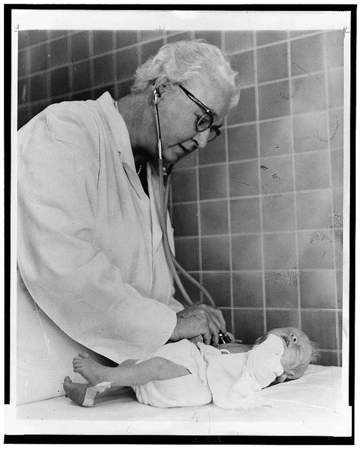
Library of Congress, Prints and Photographs Division, the New York World-Telegram and Sun Newspaper Photograph Collection, the featured photograph of Dr. Virginia Apgar examining an infant, by Al Ravenna for the World Journal Tribune, 2 October 1966, place not recorded.