Cerebral angiography began in 1927 with a needle in the artery and a radioactive dye, and for decades it was the only way to see the blood vessels inside a living human head.
A man walks out of a mine collapse with a sore neck. He talks, and his pulse is steady. His pupils react, his grip is equal, and the radiograph of his cervical spine comes back unremarkable. In 1954 that was most of what a hospital could do for him, and it could miss a torn artery entirely.
What the Film Showed
An X-ray image is made out of density. Bone stops the beam and blood does not, so a plain film of a neck shows the vertebrae in fine detail and the arteries threading past them not at all. The Museum of Radiation and Radioactivity at Oak Ridge Associated Universities states the problem in one line: to visualise blood vessels in an X-ray, it is necessary to inject a contrast agent into the blood. Without that injection there is nothing there to see. A tear in the wall of the vertebral artery, which climbs through a channel in the bone of the neck, leaves the bone unmarked. The film comes back clean and the patient is sent to observation.
Lisbon, 1927
The first pictures of living arteries inside a human head were made by Egas Moniz in Lisbon, who published the discovery in 1927 under the title L’encéphalographie Artérielle. He injected carotid arteries in cadaveric human heads and several animals before he resorted to humans, and settled first on strontium bromide, which created opaque arteries. Several attempts were necessary to obtain a successful study, and the images remained hard to interpret. The complications were real, including two patients left with Horner syndrome. And the point of it was never the artery itself. Moniz was looking for a change in the normal anatomy of the cerebral vasculature that could be indicative of a mass lesion. The vessels were a way of finding a tumour by seeing what had pushed them aside.
A Radioactive Contrast
Strontium bromide gave way to thorium dioxide, sold as Thorotrast, which remained the preferred contrast agent until the 1950s. Thorium has a very high atomic number, which is what made it a good shadow on film. Thorium is also radioactive, and it does not leave. It accumulates in the liver, the spleen and the bone marrow and stays there. The Oak Ridge museum records that between 1930 and 1955 several million individuals were injected with Thorotrast, and that a large number developed cancer, usually twenty to thirty years following the injection. Arteriography in the middle of the century was not a test anyone ordered to settle an argument. It carried a price, and a doctor weighed it against how certain he already was.
Reaching the Vertebral Artery
The carotid runs close under the skin and a needle can find it. The vertebral artery does not. It climbs through bone, and as late as 1958 a radiologist writing in the British Journal of Radiology was still calling percutaneous puncture of it a new technique, and offering an instrument to make it workable. Eric Batley listed what went wrong. The precise position of the needle point when deep in the tissues is hard to assess. The needle point is easily blunted when striking the lateral mass of the atlas, and subsequent arterial puncture is rendered more difficult. That was 1958. Four years earlier it was harder still.
October 1971
Computed tomography arrived with a single examination. Godfrey Hounsfield began developing a full-size model capable of scanning the human head in 1970, and on 1 October 1971 the radiologist James Ambrose performed the first patient scan at Atkinson Morley’s Hospital in Wimbledon, on a woman with a suspected frontal lobe tumour. Hounsfield and Allan Cormack shared the 1979 Nobel Prize for computer assisted tomography. Even that did not close this particular question. Imaging the wall of an artery rather than the channel through it came later again, and the American Journal of Neuroradiology cautions that at the site of a vertebral dissection the lumen can appear normal in calibre, with wall thickening the only sign, so the injury is easily overlooked if only lumen-opacifying studies are performed. Conventional angiography has long been considered the gold standard for these tears, and it can still miss one. No source read for this post gives the year the condition was first named.
What the Novel Takes
The Last Report gives a chapter to a night when a roof lets go underground and six men reach a company hospital at once. The one who matters is not the one screaming. He arrives flat on a stretcher talking, says he hit his neck on a timber and walked out, and his pulse is fifty-eight and steady. He is tagged green, observation. The nurse goes back to him anyway, puts her hand on his neck, and tells the physician there is a tear in the vertebral artery at C3. His X-rays show nothing, the physician says. A vertebral dissection will not show on plain film, she answers, and on that one point the record is with her. What follows is not a scan. It is rolled towels, sandbags packed flush against the jaw and shoulders, sedation, no head movement, and seven hours at the bedside waiting to learn whether the tear would lie flat. The hospital in those pages is the Copper Queen, which has a post of its own: https://duanekeelingbooks.com/blog/the-copper-queen-hospital-company-medicine-in-bisbee
The Last Report is the fourth book of The Ridge Watcher series: https://duanekeelingbooks.com/books/the-last-report
Sources
Neurocritical Care, the journal of the Neurocritical Care Society, its history of the first cerebral angiograms
The Museum of Radiation and Radioactivity at Oak Ridge Associated Universities, its account of thorium dioxide contrast agents
The British Journal of Radiology, Eric Batley, Vertebral Angiography (A New Technique), volume 31, 1958
Radiology, the journal of the Radiological Society of North America, its review Milestones in CT, and the virtual museum of the American Association of Physicists in Medicine
The American Journal of Neuroradiology, on vertebral artery dissection with a normal-appearing lumen at CT angiography
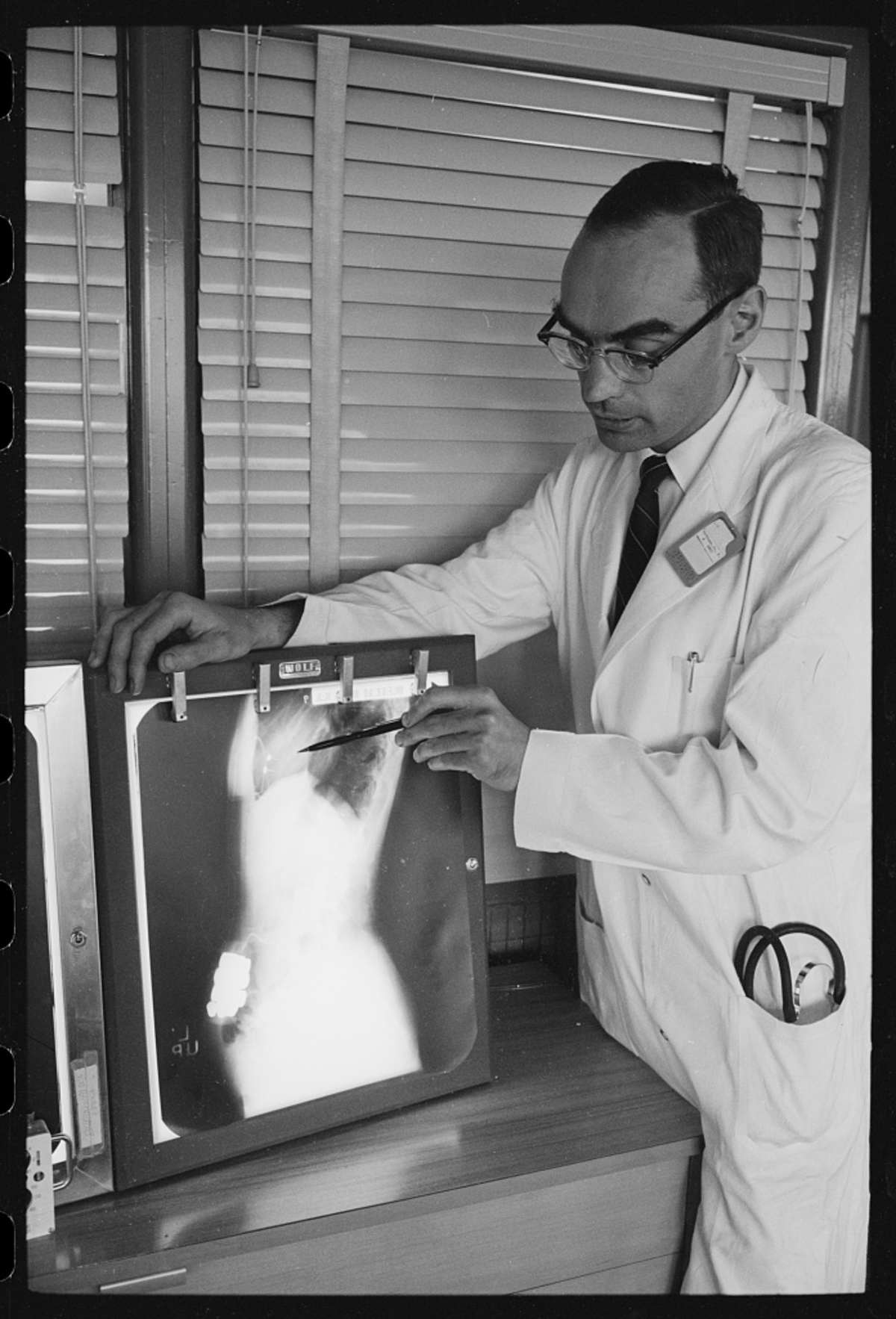
Library of Congress, Prints and Photographs Division, the U.S. News and World Report Magazine Collection, the featured photograph of a physician reading a radiograph by Warren K. Leffler, 10 May 1961, place not recorded